Original paper: Automated bone marrow cytology using deep learning to generate a histogram of cell types
Introduction
Bone marrow cytology is a critical diagnostic tool used in hematology to identify hematological disorders, such as leukemia, myeloma, and lymphoma.
However, the manual process is time-consuming, requiring trained experts to examine bone marrow aspirate digital whole slide images, and is prone to inter-observer variability, which can lead to delayed or incorrect diagnoses. An unmet need for innovative technologies to support the diagnosis of hematological disorders prompted researchers to develop an automated bone marrow cytology system.
Main Problems
How to distinguish ROI and Non-ROI patches in bone marrow aspirate digital whole slide images (WSI)?
How to identify specific types of cells on patches?
Solutions Applied
We applied DenseNet-121 to distinguish ROI and Non-ROI, and trained YOLO (You Only Look Once)v4 to identify specific types of cells on patches.
The overall process:

DenseNet-121 for ROI detection
DenseNet-121 is a convolutional neural network architecture that has shown remarkable performance on various computer vision tasks, including image classification, object detection, and segmentation. It is a popular model that has been pre-trained on a large dataset of natural images, which makes it a strong candidate for fine-tuning as a region of interest (ROI) detection model.
ROI detection is the process of identifying regions of an image that are relevant to a specific task, such as object detection, segmentation, or classification. Here, ROI detection is essential for categorizing cells in medical images.
Fine-tuning DenseNet-121 for ROI detection involves training the network on a small dataset of labeled images that are specific to the task of interest, i.e., patches with annotations of regions of interest. During training, the network is updated to optimize for the ROI classification task using the labeled data.
The advantage of fine-tuning DenseNet-121 for ROI detection is that it can leverage the pre-trained weights of the network, which have been learned on a large dataset of natural images. This pre-training provides a strong initialization for the network, which can improve the performance of the ROI detection model with a limited amount of labeled data. Additionally, the densely connected architecture of DenseNet-121 enables the network to extract features from different layers, which is beneficial for ROI detection tasks where multiple levels of features are required.
The process of region of interest (ROI) detection:
 detection model")
YOLOv4 for cell detection and classification
YOLO (You Only Look Once) is a popular object detection algorithm that can be trained to detect and classify objects in an image. YOLOv4 is a single-stage object detection algorithm, which means that it predicts bounding boxes and class probabilities in a single forward pass of the neural network. This architecture enables YOLOv4 to be fast and efficient, making it suitable for our real-time applications.
To train YOLOv4 to identify specific types of cells on patches of an image, the network is trained on a large dataset of labeled images, i.e. the patches with annotations of specific types of cells. During training, the network is updated to optimize for the cell detection and classification task.
The advantage of using YOLOv4 for cell detection and classification on patches of an image is that it can detect multiple cells in a single image patch, and classify them accurately with a high degree of confidence. Additionally, YOLOv4 has a high localization accuracy, which means that it can accurately localize the position of the detected cells in the image patch.
Another advantage of YOLOv4 is its ability to detect small objects in an image. This is particularly useful in cell detection, as cells can often be small and difficult to detect. YOLOv4 can handle small objects by using a feature pyramid network, which extracts multi-scale features from the image to detect small objects.
Applying the YOLO model to localize objects in selected region of interest (ROI) patches:

Results
The development of this automated bone marrow cytology system has significant potential to support more efficient and accurate diagnoses in hematology. The results of the study demonstrated that the system achieved high accuracy in region detection, cell detection, and cell classification. The system achieved an accuracy of 0.97 and a ROC AUC of 0.99 in region detection. The mean average precision and average F1-score were 0.75 and 0.78, respectively, with a log-average miss rate of 0.31 in cell detection and classification.
The technology can assist pathologists in achieving more accurate diagnoses and treatment plans, ultimately benefiting patients. The automated system has the potential to reduce the time and cost associated with manual bone marrow cytology, making the diagnosis more accessible to patients in rural and remote areas.
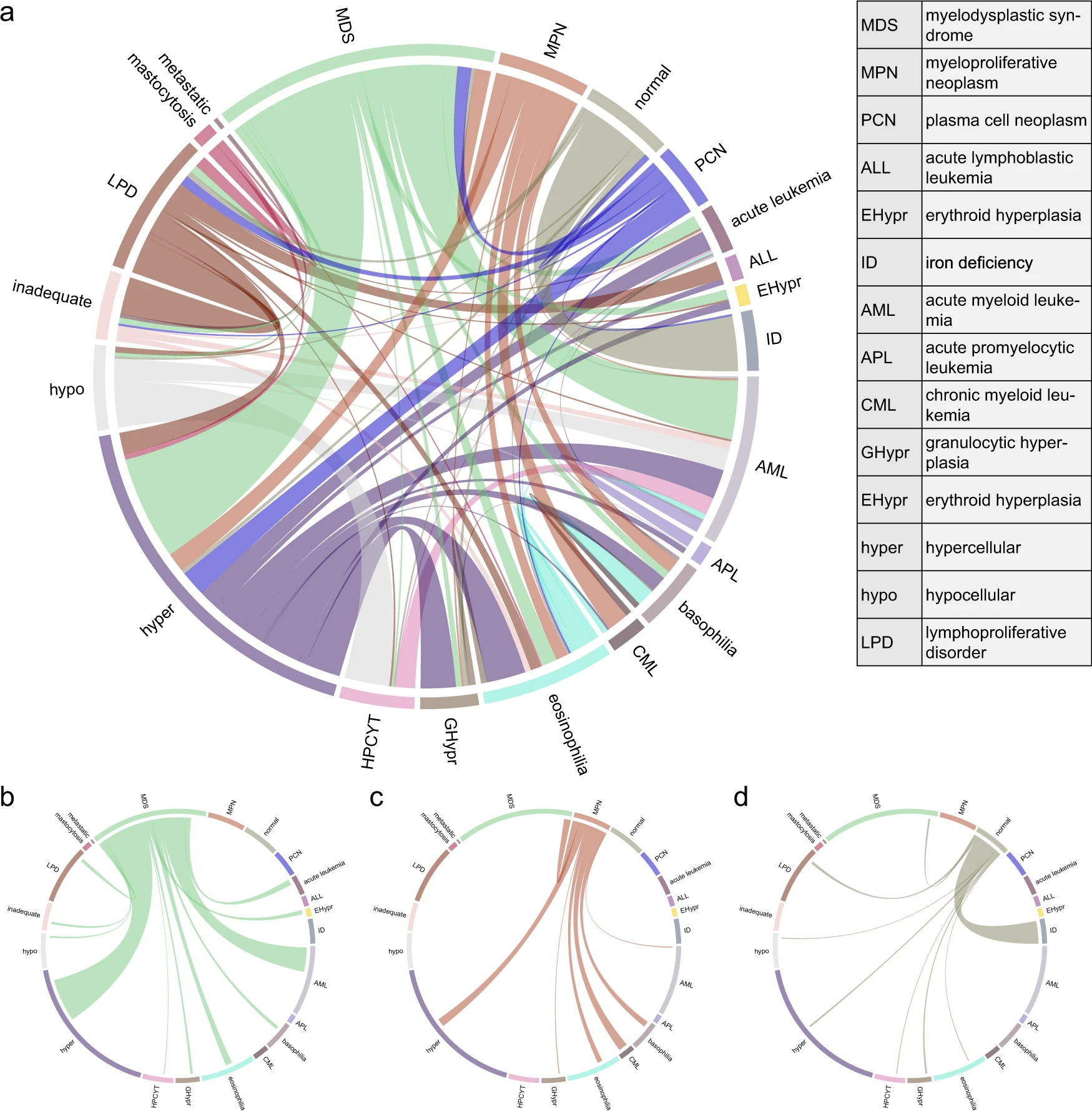
Generating the Histogram of Cell Types (HCT) and converged Integrated Histogram of Cell Types:
 and converged Integrated Histogram of Cell Types")